Field of Science

Showing posts with label physiology. Show all posts

Anti-clotting factors in vampire bat saliva may save your life.

While I'm on the subject of things that suck blood, I'd like to take a moment to tell you how vampire bat saliva may save your life one day.

Yesterday I happened to see a tweet from the Ohio State University Medical Center's twitter account that linked to a press release discussing a new plasminogen activator that is currently undergoing clinical trials. The drug, desmoteplase, is modeled after a protein found in vampire bat saliva that prevents clots and platelet aggregation, which keeps the blood flowing while the bat is feeding. Plasminogen activators like desmoteplase are used to break down blood clots that are blocking blood flow to vital organs such as the heart, lungs, or brain.


Anatomy of a heart attack.
Image credit: Flickr user gandhiji40
To understand why bats have this protein in their saliva and why it may be medically useful, we first need to understand how clots form. Whenever a blood vessel is damaged, collagen fibers are exposed under the severed lining of the vessel. Platelets, one of the several blood cell types, will begin to stick to the collagen fibers and to one another until the damaged area is covered. This creates a platelet "plug" that stops immediate blood loss. Once the platelet plug is in place, a cascade of clotting factors (a vital one being a chemical called fibrin) will build a clot "seal" over the platelet plug that forms a more permanent barrier to blood loss while the vessel heals.

After the vessel heals and the clot is no longer needed, a chemical called plasminogen is activated and becomes plasmin. Plasmin then breaks down the clot by solubilizing fibrin. This is a safe way to get rid of the clot so that it doesn't come off in one piece and then lodge itself into a small vessel where it can cut off circulation to parts of the body. However, sometimes clots are not broken down properly and can cut off blood flow to the brain (stroke) or heart (heart attack). When this happens, we need to manually turn plasminogen into plasmin using plasminogen activators, so that plasmin can break down the clot and restore blood flow.

Vampire bat, Desmodus rotundus.
Image credit: Flickr user Robertsphotos1
The plasminogen activators in vampire bat saliva were first described by Dr. Christine Hawkey in a letter to Nature in the 1960s. In addition to plasminogen activators, Hawkey later went on to describe platelet aggregation inhibitors in vampire bat saliva as well. Vampire bats do not suck blood directly as mosquitoes do; instead they puncture the skin with their teeth and lap the blood with their tongues as it seeps through the wound. This strategy requires the inhibition of platelet plugs and clots which would stop the blood from continuing to flow during the bat's meal.

Desmoteplase, which is derived from the plasminogen activators in vampire bat saliva originally described by Hawkey, seems to have some advantages over currently used plasminogen activators (which are often based on chemicals in humans). Current drugs are only approved for use up to 3 hours after symptom onset, and Dr. Michel Torbey at OSU MC is hopeful that desmoteplase will demonstrate efficacy up to 9 hours after symptom onset, which could drastically reduce the number of deaths. From the press release:

"Prompt medical care within three hours is very important for recovery from a stroke, but attempts to find drugs that extend the treatment window have not been successful," added Torbey. "If the study findings back up our hopes and expectations, desmoteplase could be a real game changer in our ability to help patients."

In addition to expanding the treatment window, desmoteplase is more potent and specific than current drugs. One current plasminogen activator is even linked to neurotoxicity in some patients, so there is high demand for newer and better drugs to treat problematic clots. If approved, this drug could reduce the risk of death in stroke patients who live in remote areas and may not be able to make it to the emergency room within the three hour window.


ResearchBlogging.orgHAWKEY, C. (1966). Plasminogen Activator in Saliva of the Vampire Bat Desmodus rotundus Nature, 211 (5047), 434-435 DOI: 10.1038/211434c0

Hawkey, C. (1967). Inhibitor of Platelet Aggregation Present in Saliva of the Vampire Bat Desmodus rotundus British Journal of Haematology, 13 (6), 1014-1020 DOI: 10.1111/j.1365-2141.1967.tb08870.x

Schleuning, W. (2001). Vampire Bat Plasminogen Activator DSPA-Alpha-1 (Desmoteplase): A Thrombolytic Drug Optimized by Natural Selection Pathophysiology of Haemostasis and Thrombosis, 31 (3-6), 118-122 DOI: 10.1159/000048054

Heat shock proteins keep mosquitoes fertile after they feed on you.

One of my committee members is an insect physiologist. I picked him for my committee because he's an excellent physiologist who has experience with lipid chemistry. (He's also an all around nice guy and a very successful scientist.) I took a photoperiodism seminar that he taught, and it was my favorite seminar class in graduate school. Despite all of this, my understanding of insect physiology is... almost nonexistent. I know a little bit about diapause from his seminar and a writing class that I took with one of his advisees, but that's about it. I'd like to learn, and if I had another year or two of grad school ahead of me I'd probably take an insect physiology class. As a wise man once said, "If 'if's and 'but's were candies and nuts then we'd all have a Merry Christmas," am I right?

Anyway, my point is that he has a paper in PNAS this week that I found really interesting, but you'll have to forgive me if there are any physiological inaccuracies in this post. I claim responsibility for anything that's wrong.


Aedes aegypti. Credit: Flickr user Sanofi Pasteur
It may not seem like it during this part of the year (in the northern hemisphere anyway), but most of the time, mosquitoes don't drink blood. Males and females both drink nectar for their own survival; it is the propagation of the species that requires your blood. Female mosquitoes sometimes need to take a hot blood meal to get the required proteins and iron for making eggs. While necessary for reproduction, drinking mammalian blood has a lot of unique physiological challenges, not the least of which is the temperature difference.

At room temperature, the average human's body temperature is about 15°C (almost 30°F) warmer than that of the average mosquito, and when a female takes a blood meal, her body temperature spikes 10°C in one minute! While ectotherms like mosquitoes are used to fluctuating body temperatures based on environmental conditions (such as the day/night cycle), these changes are usually gradual, allowing the mosquito ample time to alter their physiology. Hot blood meals impose the unique physiological problem of rapidly increasing body temperature without much time to adjust, which can cause enzymatic dysfunction and disrupt physiological pathways such as digestion, reproduction, and metabolism.

Obviously mosquitoes are not dying out because they are heat stressed and can't reproduce (wouldn't that be great?), so how are they getting around this problem? Heat shock proteins! Heat shock proteins can aid as enzymatic "chaperones", making sure that the enzyme proteins stay folded in the proper conformation during times of physiological stress (not just heat but also extreme cold, lack of oxygen, infection, or exposure to toxins, among other things). Enzymes are a key player in digestion; they aid in the breakdown of proteins, fats, and carbohydrates, and the absorption of certain nutrients, so the mosquitoes definitely want to conserve their function right after taking a meal.

It turns out that female mosquitoes experience an 8-fold spike in heat shock protein expression in the midgut following a hot blood meal. When the mosquitoes were prevented from being able to express HSP70 (the heat shock protein active in these mosquitoes), the blood proteins from their meal stayed in the midgut longer, suggesting that digestion of blood proteins is somehow impaired. It is unclear whether this is due to a deficiency in breaking down the proteins or slower uptake of proteins and nutrients from the midgut, but the result is the same: the proteins are staying in the midgut longer instead of going into the body and aiding egg formation.

While heat shock proteins are not preferentially expressed in the ovaries after a hot blood meal, the mosquitoes prevented from expressing HSP70 also made fewer eggs than control mosquitoes, which provides further evidence that the delay of protein digestion and nutrient absorption is interfering with egg production. Since mosquitoes are vectors for many human diseases, research like this can be used towards initiatives to potentially limit the fecundity of mosquitoes (i.e. how rapidly they reproduce) and reduce human infection with diseases like yellow fever, malaria, and dengue fever.


ResearchBlogging.orgBenoit, J., Lopez-Martinez, G., Patrick, K., Phillips, Z., Krause, T., & Denlinger, D. (2011). Drinking a hot blood meal elicits a protective heat shock response in mosquitoes Proceedings of the National Academy of Sciences DOI: 10.1073/pnas.1105195108

Total calorie intake is most influential in regulating adiponectin

Today I have a guest post over at the LabSpaces guest blog Dangerous Experiments. In that post I discuss a recent paper that examines the relative influence of total caloric intake, relative amount of dietary fat, and existing body fat on the circulating levels of an endocrine called adiponectin. Adiponectin is involved in energy homeostasis, specifically glucose uptake and the breakdown of fat, among other things. Hypoadiponectinemia (having too little adiponectin) is a risk factor for a lot of obesity-related diseases, like Type II Diabetes and metabolic syndrome.


ResearchBlogging.orgLiping Qiao, Bonggi Lee, Brice Kinney, Hyung sun Yoo, and Jianhua Shao (2011). Energy intake and adiponectin gene expression Am J Physiol Endocrinol Metab. : 10.​1152/​ajpendo.​00004.​2011

A question for psych/neuro/gastro friends: walking, anxiety, and emesis.

I may or may not have mentioned before on this blog that I have a specific phobia of vomiting. If I haven't, now you know. I don't like to talk about it in my online life because it is a major source of stress in my offline life (it is the root of the agoraphobia that I know I have mentioned), but I bring it up today because I have some unanswered questions that I'd like to put out into the blogosphere.

For those of you who are not familiar, I will try to outline the nature of emetophobia, at least the way I experience it. It stems ultimately from a fear of losing control of one's body. Vomiting is unavoidable, violent, and sometimes unpredictable. It isn't fun for anyone (well, almost anyone), but for an emetophobe it becomes a life or death situation. That loss of control is not just unpleasant, it is entirely unbearable and intolerable, and the mere thought of losing control in such a manner is enough to arouse panic and hysteria. As a result, an emetophobe becomes hyperalert to internal and external cues that vomiting is about to happen. Internal cues like nausea, or external cues like someone else actually vomiting, saying they don't feel well, saying they had vomited recently, etc. It differs from person to person, and some people are only afraid of themselves vomiting whereas others are afraid of themselves and others vomiting. To speak for myself, the only reason why I'm afraid of someone else vomiting is because I'm afraid I'll catch whatever they have and, as a result, vomit myself.

As a result of this state of being hyperalert, there are undoubtedly a lot of false positives. Emetophobes are notorious for not being able to decipher their own gastrointestinal cues. Non-pathological sensations like having gas or feeling full are misinterpreted as nausea. Nausea (real or perceived) causes anxiety, and one of the symptoms of anxiety is, yeah, more nausea. It becomes a terrible positive feedback loop that can be really hard to escape once the ball is rolling.

I have several coping mechanisms that I use when I feel the anxiety beast starting to swell inside me. They fall into several categories: preparative, preventative, and distractive. Preparative behaviors are things that will ease the process if I actually do throw up, such as taking off loose clothing, tying back my hair, and making sure I have access to a toilet. Preventative measures are things like taking antiemetics, benzodiazepines, sipping water, sucking on peppermints, etc. The distractions, however, seem to be the most effective.

My distraction techniques usually have two horns: giving my body a menial task to accomplish and giving the rest of my brain something to focus on. Generally this involves walking around my apartment and either having a conversation with my boyfriend (if he's around and willing to oblige) or putting something light and fluffy on the TV. I'm fond of sitcoms and baking shows (like Cupcake Wars) for this situation. The TV/conversation part is easy to understand: I'm providing an easy distraction for my brain to focus on instead of thinking about the nausea and anxiety. If I don't focus on it, most of the time it will go away on its own. The walking part is less easy for me to understand.

I know that having a menial activity to focus on is part of the distraction process. If I'm focused on walking around and balancing, even if only a tiny part of my brain is involved, that's one less bit of my brain that can be thinking about my stomach. But, that's not the only thing. The walking actually jostles my body, and adds "noise" to the internal cues that I'm misinterpreting as nausea. The walking seems to literally settle my stomach by covering up the stimulus with other sensations. But finally, I can't fully rule out a physiological connection. Could there be a chemical reason why walking settles the stomach or reduces anxiety? I've heard of "walking through the pain", but can you walk through the fear as well? I don't know enough psychology or neuroscience (I imagine the brain-gut axis is very important in this response, but I don't know how) to know the answer to that, so I'm hoping that someone out there will. Can physical movements calm the sympathetic response?

As a side note, I have often wondered if there was a physiological reason why emetophobes claim to vomit less often than normal people. I know that the sympathetic response causes gastrointestinal motility to slow down. If fear slows down peristalsis, could it also slow down reverse peristalsis?

[Image credit: Lauren Brooke. I had to search for a good image with my hand over my face, peeking through my fingers. Shockingly I was spared from having to see any images of actual people actually vomiting. Score one for me.]


ResearchBlogging.orgBOSCHEN, M. (2007). Reconceptualizing emetophobia: A cognitive–behavioral formulation and research agenda Journal of Anxiety Disorders, 21 (3), 407-419 DOI: 10.1016/j.janxdis.2006.06.007

Davidson, A., Boyle, C., & Lauchlan, F. (2008). Scared to lose control? General and health locus of control in females with a phobia of vomiting Journal of Clinical Psychology, 64 (1), 30-39 DOI: 10.1002/jclp.20431

Lipsitz, J., Fyer, A., Paterniti, A., & Klein, D. (2001). Emetophobia: Preliminary results of an internet survey Depression and Anxiety, 14 (2), 149-152 DOI: 10.1002/da.1058

Veale, D., & Lambrou, C. (2006). The Psychopathology of Vomit Phobia Behavioural and Cognitive Psychotherapy, 34 (02) DOI: 10.1017/S1352465805002754

What's the deal with male circumcision and female cervical cancer?

Today I have a post over at the Scientific American Guest Blog on male circumcision and cervical cancer. In the post I discuss several papers on the efficacy of circumcision in reducing cervical cancer risk, the physiology behind how circumcision slows the spread of human papilloma virus, and the arguments against circumcision as a prophylactic.


Recently, while I was getting drinks at a pub with about a dozen or so other biologists, I was involved in a very animated discussion about circumcision -- because that's what biologists argue about when they're drunk, apparently.

"They do it to increase stamina. It desensitizes the penis," said a microbiologist. (There's some evidence to the contrary on the bit about stamina, actually.)

"Actually there are studies that show that circumcision decreases the risk of cervical cancer," added an entomologist.

"I have a foreskin and I'm proud of it. I promise you none of my partners have cervical cancer. I think it has more to do with whether or not they practice good hygiene," said an animal biologist (thanks for sharing that with us, by the way). By this point we were starting to get funny looks from the other patrons, and the conversation dissolved without much consensus as to whether circumcision inherently prevents the spread of HPV and cervical cancer, or if not being circumcised merely compounds the issue of poor hygiene. Being the good scientist I am, I decided to dip into the literature to get to the root of the discussion. For now, let's ignore the debates over whether or not circumcision reduces genital sensitivity or the spread of HIV, although these are certainly important things to consider when deciding whether or not to circumcise your children.

Does male circumcision reduce the risk of cervical cancer in their female partners?

A 2002 paper in the New England Journal of Medicine studied men in Europe, Asia, and Latin America, and found that circumcision was correlated with a decreased risk of penile HPV infection (this correlation is corroborated by a 2009 study in African men), but that there was not a significant correlation between circumcision and incidence of cervical cancer.

When they restricted their dataset to women with only one sexual partner, there was an increased risk of cervical cancer in women whose partners were uncircumcised only if their partner was already considered at high risk for contracting HPV (as determined by age at first intercourse, number of sexual partners, and sex with prostitutes). So, in men who already engage in risky sexual behavior, circumcision does offer an advantage for protecting their partners from cervical cancer.

A more recent paper published this year in The Lancet studied HIV negative men and their partners in Africa, circumcising half of the men immediately and the other half after 2 years. Their female partners were tested for high risk genotypes of HPV that are known to cause cervical cancer at the beginning of the study, as well as 1 year and 2 years afterwards. After controlling for lifestyle variables, the women partnered with men who were circumcised had significantly reduced rates of infection with both low and high risk HPV genotypes. However, the women in this study were overwhelmingly monogamous (only 4% of female participants had more than one sexual partner in the year prior to the study), so the results cannot be extrapolated to women with multiple sexual partners.

How does circumcision offer an advantage in reducing the risk of HPV?

Most of the penis is covered by keratinized epithelium that is typical of most other parts of the body. Keratinized epithelium, which has an outer layer of dead cells called the stratum corneum, offers many advantages for protection against viral infections. It is more durable and less likely to tear, so it offers a physical barrier between pathogens and the inside of the body, as well as various chemical defenses against infection and dead outer layers that are constantly being shed, taking any external pathogens with them. However, the inner foreskin has a mucosal lining that is not keratinized, therefore more prone to minute tearing and infection. This mucosal inner surface is pulled back and exposed during intercourse and made susceptible to the transmission of HPV and other viruses. Circumcision reduces the mucosal surface area, thereby potentially minimizing the interface for abrasion and transmission of viruses.

So then what's the problem here? What are the other viewpoints?

From the letters in response to the 2002 study [PDF]:

"Human papilloma virus causes cervical cancer; the foreskin does not. Safer sex, not circumcision, prevents the spread of HPV," says George Hill of Doctors Opposing Circumcision.

"Because infants are not sexually active, they should not be required to bear the bur­den of preventing sexually transmitted infections. Sexually transmitted dis­eases will be prevented by practising safer sex, not by circumcising infants. If circumcision is touted as a prophy­lactic, it could confer a false sense of security and encourage high-risk sex­ual behavior," writes Arif Bhimji and Dennis Harrison of the Association for Genital Integrity.

While there is evidence to show that circumcision offers an advantage in preventing cervical cancer, it is by no means a cure. The number of sexual partners that individuals may have is a confounding variable, as increased partners or risky sexual behaviors result in an increased incidence in HPV and cervical cancer across all groups. In addition, while it is true that women with circumcised partners are less likely to get cervical cancer, they are not immune. Women with circumcised partners still contract HPV and develop cervical cancer! They just do it at a reduced rate. There are other methods that are much more likely to reduce a woman's chance of contracting HPV and developing cervical cancer, such as vaccination and condom use. Therefore, from a public health standpoint in the United States, it may not be necessary to circumcise male babies solely for the purpose of reducing the risk of cervical cancer in his future sexual partners (of course, this doesn't take into account the possibility that the child might not be heterosexual). This would also be true in similar societies where there is sufficient access to education on safe sexual practices, condoms, and HPV vaccinations. However, the decision whether or not to circumcise a child is a complex one involving cultural, religious, health, and geographic variables, just to name a few.

As for my friend the animal biologist, I shared these papers with him and he came to the same conclusion that I did (though in slightly more colorful language). As long as you and your partner are willing and able to practice safe sex, an uncircumcised penis isn't any more likely to give you cervical cancer than a circumcised one.

Originally published by Scientific American, Inc.


ResearchBlogging.orgCastellsagué, X., Bosch, F., Muñoz, N., Meijer, C., Shah, K., de Sanjosé, S., Eluf-Neto, J., Ngelangel, C., Chichareon, S., Smith, J., Herrero, R., Moreno, V., & Franceschi, S. (2002). Male Circumcision, Penile Human Papillomavirus Infection, and Cervical Cancer in Female Partners New England Journal of Medicine, 346 (15), 1105-1112 DOI: 10.1056/NEJMoa011688

Wawer, M., Tobian, A., Kigozi, G., Kong, X., Gravitt, P., Serwadda, D., Nalugoda, F., Makumbi, F., Ssempiija, V., Sewankambo, N., Watya, S., Eaton, K., Oliver, A., Chen, M., Reynolds, S., Quinn, T., & Gray, R. (2011). Effect of circumcision of HIV-negative men on transmission of human papillomavirus to HIV-negative women: a randomised trial in Rakai, Uganda The Lancet, 377 (9761), 209-218 DOI: 10.1016/S0140-6736(10)61967-8

What "I Love Lucy" can teach us about capillary nutrient exchange

During yesterday's review session we went over the circulatory system, an important part of which is the exchange of nutrients, gasses, and waste products between the blood and the body's tissues at the capillaries. Nutrients and gasses diffuse into the tissues to be used for cell proliferation, metabolism, and other things, and the waste products are taken up by the blood to be removed at the lungs or kidneys.


(I lost the source for this image. If it is yours, contact me and I'll credit you.)

We maximize nutrient exchange by increasing the surface area for diffusion (lots of tiny vessels instead of one big vessel) and slowing down the blood once it gets to the capillaries so it spends more time in the tissues. The professor for the class originally explained nutrient exchange as someone trying to pick candy off of a conveyor belt. We want the conveyor belt to move slowly so we can maximize the number of candies we can grab off of it before they pass us by. I thought this was a clever way to explain the concept, because my mind immediately went to the classic chocolate factory scene in I Love Lucy.

However... in the office hours and at the review session it became increasingly apparent to my co-TA and I that many of our students weren't getting the reference. And since they didn't get the reference, they didn't really understand the analogy. We told them to look it up on Youtube when they got home, but one student had his laptop in the room so he looked it up immediately. Other students watched it over his shoulder... began to laugh... and I could see the click happen. Suddenly they got it.


Seriously, what kind of person has never seen the chocolate factory episode of I Love Lucy? I didn't think there was a generation gap between my students and I, but now I'm starting to wonder.

Weird sex physiology, Halloween edition


Source. Myotis lucifugus. © M. A. Tuttle, Bat Conservation International

ResearchBlogging.orgOne physiological problem (of many) that hibernating mammals face is the preservation of gametes during the winter months. During torpor, most animals do not continue spermatogenesis (the creation of new sperm) because it would require too much energy, but they also need sperm to be immediately available when they awake. This is because in many cases, the earliest born offspring have the best chance of surviving the following winter because they have more time to develop and build energy stores. So, hibernating animals must find a way to preserve the sperm they make during the period leading up to the onset of torpor.

Several bat species, such as the Myotis lucifugus pictured above, have evolved an interesting way to combat this issue: they increase the osmolality (the ratio of solutes to water) in the epididymal tubes where the sperm are stored. This works for three reasons. First, the epididymal tubes are deeper in the body than the testes, so storing the sperm there will reduce the drop in temperature experienced by the sperm. Second, the increase in extracellular solutes draws water out of the sperm cells, making them immobile. Third, the increase of solutes in the extracellular fluid reduces the activation energy of water, affording the cell membranes extra resistance to "cold shock".


Fig 1: Model of events in bat epididymis prior to torpor (adapted from Critchton et al., 1994).

To model how this process might happen, various solutes enter the epididymal tubes, drawing water in with them via osmosis and causing the epididymal tubes to swell. The solutes are then converted into forms that are impermeable to the epididymal epithelium and the sperm plasma membrane, so that water stays in the epididymal tubes and extra water is drawn out of the sperm cells, which shrink and become immobile until the end of hibernation. In some species of bats, the epididymal osmolality can be five times larger than plasma osmolality, and 3-4 times larger than normal epididymal osmolalities during active months!


Fig 2: Electron micrographs of bat spermatozoa at different osmolalities. 
A: 100 mmol/kg water, B: 300, C: 500, D: 1000.

The first impression I get from this picture is, wow, bat sperm is kinda scary looking and seems very appropriate for a Halloween post. But back on topic: bat sperm seems to be most active between the osmolalities shown in panels B and C, where D is osmolality during hibernation, and A is when the sperm cells begin to lyse (burst) from the influx of water due to hypertonicitiy (you can see the plasma membrane of the head of the one sperm cell beginning to swell in this photo).

The epididymis of bats normally has a slightly higher osmolality than the blood plasma, and it appears that these bat species have exploited this condition to the extreme during their hibernating months to maintain their vitality upon wakening.


Happy Halloween everyone!


E. G. Crichton, B. T. Hinton, T. L. Pallone and R. H. Hammerstedt (1994). Hyperosmolality and sperm storage in hibernating bats: prolongation of sperm life by dehydration Am J Physiol Regul Integr Comp Physiol, 267, 1363-1370

Pictures and video for a Sunday afternoon.


Source. A reader pointed me to a second composite image of a "global human." It is "a composite itself from four composite of Northwest European, South & West Asian, East Asian and African faces..."

As I said in the comments at Razib's blog: "My, that is one attractive fellow. He looks decidedly Hispanic to me, which I suppose shouldn't be surprising since Hispanics are already highly mixed people with Asian, African, and European ancestry. However, if you squint, he can look a bit Indian (or Italian, or mulatto, or hapa, or…)." I think the thought exercise here is less what the guy actually looks like, but more what we as individuals see in him.


Source. To correct [people's misinterpretations about the relative size of Africa due to maps using the Mercator projection], Kai Krause has created this map and associated information to show that Africa's total area is almost the same as that of the United States, China, India, Japan and all of Europe combined.

I learned about the faults of Mercator projections when I was pretty young, although I can't remember where I learned it. I think it was something I saw on television. My young brain couldn't quite wrap itself around the geometry involved, but I got the major point. Many maps use a system that, instead of staying true to the curvature of the earth, lay things out in such a way that the lines of latitude and longitude make rectangles instead of curved lines. This is mainly for ship navigation and the like, but it has the unfortunate effect of exaggerating land masses at the poles and diminishing land masses near the equator, such as central America, Africa, and India. In the Mercator projection, Africa looks to be only slightly larger than Greenland! If you've ever looked at a globe (I had a globe when I was a kid, which helped me with the geometry of this issue a bit), you know that's definitely not true. The original blog post has more detailed info on Mercator projections if you're interested.


Source. Myotonia congenita, or congenital myotonia is a skeletal muscle-locking disorder. In affected individuals, when the muscles contract, they do not immediately relax again: for a short period the affected muscles stiffen. If the individuals are standing up, they'll fall over, as the kittens in the video do.

I felt bad watching this video. Every time a kitten would fall down, my immediate instinct was to laugh, but then I wanted to cry. I had a very conflicting experience to say the least. The blog post accompanying this video has a really great explanation of the physiology involved in the disease; I suggest you check that out as well.

Dick Hill: Stinky Clams and Coral Bleaching

ResearchBlogging.orgYesterday I had the opportunity to chat with Dick Hill, the author of one of the most popular animal physiology textbooks in circulation right now, and a pretty damn successful ecophysiologist. He used to work with mammals and birds, but now he's moved on to marine invertebrates, and he's currently studying betaines as a possible defense mechanism against coral reef bleaching.

He got into coral bleaching as a bit of a happy accident. He was doing work on giant clams in the south Pacific, investigating why the giant clams took on a foul odor hours after being killed. The locals had begun a major operation to farm and sell the giant clams around the world as delicacies (apparently they're very delicious), but even when frozen, the clams were inedible after a few hours because of the breakdown of a chemical called dimethylsulfoniopropionate (DSMP). The clams are symbionts with a species of algae that produce very large quantities of DSMP, and as a result the DSMP settles in their bodies in quantities that are orders of magnitude larger than similar marine animals. After the animal dies, the DSMP breaks down, and one of the by-products is dimethylsulfide, which accounts for the rank odor of the clams. The happy accident comes in when he began using mass spectrometry to affirm the presence and by-products of DSMP in the clams. One of the mass spectrometry specialists that he worked with told him that there were multiple types of betaines in the clams as well!

Betaines are well known to exist in various plants, especially crop plants, many of which have been genetically engineered to express more betaines. From my understanding, betaines can act as osmolytes (compounds that affect the diffusion of water through membranes) and help protect membranes and proteins from various stressors. In plants this generally means protecting photosynthesis pathways from high temperatures and high levels of irradiation from the sun during the peak of the daylight cycle. You would think that more sunlight would be good for photosynthesis, but apparently it is the opposite because of the negative effect of irradiation. As a result there is often a dip in photosynthesis productivity in the afternoon (a phenomenon called photoinhibition) unless the plant can shield itself from harmful irradiation using betaines.

This information is important to corals because of the decline in coral populations due to bleaching. 'Bleaching' occurs when corals lose their algal symbionts (which are what give corals their brilliant colors) due to high water temperature and light intensity. When the symbionts leave or die due to photoinhibition, it is more difficult for the corals to survive. While corals were known to contain betaines for osmotic protection, the possible role of betaines in the stabilization of photosynthesis pathways in corals had been largely ignored until recently. With this in mind, Hill and colleagues set out to determine ecological patterns in coral betaine concentrations, which would be quite valuable for informing future conservation efforts.

Hill found that colonies of corals at shallower depths had higher concentrations of betaines than colonies in deeper waters and that colonies living in exposed areas had higher concentration of betaine than shaded colonies. This is very good evidence (although not proof of causation) that betaines are suppressing photoinhibition in corals, because exposed colonies and colonies in shallower water experience higher water temperatures and levels of irradiation than their deeper or shaded counterparts. In addition, they found that betaine levels were higher in the afternoon than in the morning, suggesting that betaine levels were susceptible to phenotypic plasticity in response to more direct sunlight!

Hill is currently investigating the function of the betaines in these corals, to determine whether or not they are actually stabilizing photosynthesis in corals as they do in vascular plants. He is also investigating whether or not the betaines are being produced by the corals themselves, or by the algal symbionts, or both.

[Please feel free to correct me if I've misinterpreted anything; I will freely admit that I'm a tetrapod lady, and I know little about plants and invertebrates!]


Hill, R., Dacey, J., Hill, S., Edward, A., & Hicks, W. (2004). Dimethylsulfoniopropionate in six species of giant clams and the evolution of dimethylsulfide after death Canadian Journal of Fisheries and Aquatic Sciences, 61 (5), 758-764 DOI: 10.1139/f04-029
Hill, R., Li, C., Jones, A., Gunn, J., & Frade, P. (2010). Abundant betaines in reef-building corals and ecological indicators of a photoprotective role Coral Reefs DOI: 10.1007/s00338-010-0662-x

Neuromuscular Junction Cookie!

This is adorable. From here, found via this science cookie roundup post that I also highly recommend you visit, if you enjoy stuff like anatomical cakes and cookies that resemble T-4 macrophages.


Click to enlarge!

On detecting stress endocrines in hamster poop

I realize I am cutting in just under the wire, but for today's rodent blogging, I'd like to discuss three things that are very near and dear to my heart: hamsters, stress endocrines, and poop.

This wonderful little specimen is a Syrian (or golden) hamster:


ResearchBlogging.orgThere is some evidence to show that hamsters use cortisol as a primary stress endocrine, similar to humans, which would make them a better model for studying stress responses that can be extrapolated onto humans, as opposed to rats and mice, which primarily use corticosterone. There is some debate about the extent to which hamsters use coristol, and both cortisol and corticosterone are present in hamsters, so researchers are currently duking that issue out. In any case, corticosterone is a precursor to cortisol, and both are involved in the stress endocrine cascade, which I have blogged about before.

Traditionally in such hamster stress models, measurements of plasma corticosteroids were taken from the blood, and the preferred methods of getting enough blood to do so were fatal to the animal (I will spare you the gory details, but I'm sure you can imagine). You can't take multiple measurements over time on a dead animal, so in their paper, Sex differences in the excretion of fecal glucocorticoid metabolites in the Syrian hamster, Chelini et al. investigate an alternate, much less invasive way to sample stress endocrines: by looking at their poop!

Poop is easily collected without disturbance to the study animal and can be collected many times as time progresses, which is a good thing, but the metabolism and excretion of different stress hormones are different, and can differ between animals, primarily due to sex, so Chelini and colleagues designed an experiment to attempt to validate this method. To do this, they injected hamsters with adrenocorticotropic hormone (ACTH), which is the endocrine released by the anterior pituitary to tell the adrenal cortex to start making stress endocrines (more in that in this post), and a control group received a saline injection. They collected feces in four hour intervals one day before and two days after injection, then extracted (I'm really glad I'm not the person whose job it was to vortex all those poop samples) and quantified the stress endocrine metabolites present in the feces.


Fig. 1 Variation of fecal glucocorticoid metabolites (FGM) over 24 h in male (n = 20; white boxes) and female (n = 21; light gray boxes) Syrian hamsters

The first thing they found, during the 24 hours pre-ACTH injection, was that male hamsters had more fecal glucocorticoid metabolites (FGM) than females. Male baseline measurements were about four times higher than the concentrations of FGM found in females. These concentrations differed throughout the day in different ways between males and females as well.


Figs. 2 & 3 Boxplots of fecal glucocorticoid metabolites (FGM; ng/g) of male [and female] Syrian hamsters after an i.m. injection of adrenocorticotropic hormone (ACTH; dark gray boxes) or saline solution (Control; white boxes). Respective median baseline values of the ACTH (filled circle with broken lines) and control group (open circle with dotted lines) at the same time of the day are also shown. Asterisks beside the box indicate significant differences compared with the baseline levels of the same group

Compared to controls and pre-injection measurements, the experimental group that received the ACTH injection had elevated FGM in their feces after about 8 hours post-injection in both males and females. Males experienced a spike in FGM eight hours earlier (20 hours post-injection) than females, and still had elevated levels of FGM after 48 hours, whereas females were back to baseline by that time.

The successful validation of these methods is important because, since individuals can be sampled multiple times, it allows them to serve as their own controls. Differences between individuals can vary up to ten-fold in FGM concentration! Their methods also reflect the natural sexual dimorphism in these hamsters: males have larger adrenal glands and secrete more corticosteroids than females. Interestingly, the injection procedure itself did not cause a significant rise in FGM in male controls, but it did in female controls (~32 hours post-injection, around the same time as the peak FGM of experimental females). This would imply that the injection procedure is more stressful for female than male hamsters. I don't have a good idea offhand as to why that may be.

In conclusion, our results show for the first time in the Syrian hamster that adrenocortical activity can be monitored in fecal samples in a non-invasive way. Actually, the satisfactory results generated by our [methods] demonstrated clearly that changes in glucocorticoid concentrations in the blood are well reflected in changes in FGM in both sexes. The difference we observed between male and female FGM levels are in accordance with the sex difference reported by others regarding the hamster adrenal physiology. Therefore, our study provides a humane, practical, and noninvasive alternative to blood removal, and therefore a powerful tool for stress-related studies in a species where venepuncture has traditionally been regarded as problematic. This is especially important, as hamsters are used as an animal model for medical research.
I, for one, strongly approve of the development of non-invasive, non-lethal sampling methods for fuzzy things. I mean, c'mon, look at this guy:


Chelini, M., Otta, E., Yamakita, C., & Palme, R. (2010). Sex differences in the excretion of fecal glucocorticoid metabolites in the Syrian hamster Journal of Comparative Physiology B, 180 (6), 919-925 DOI: 10.1007/s00360-010-0467-9

SHARK WEEK!

In case you hadn't heard, it's Shark Week, an annual week-long event by The Discovery Channel that showcases everyone's favorite ocean predator. And this year Michelle is here to teach you a little bit about shark physiology. In the interest of full disclosure, sharks are not my forte. I am a tetrapod girl, but I know just enough about sharks to know that they have a really, really cool electrosensory system that helps them catch prey.


Most people know that sharks have an acute chemosensory system that enables them to detect very dilute amounts of blood in the ocean water, but the ability to 'smell' blood only gets a shark so far. Once the shark gets closer to its prey, the chemical trail left by the blood floating in the water is no longer accurate. The prey, now alert to the shark's presence, can dart around and distort the signal by distributing blood in all directions. The shark now needs to rely on some other sense, and vision is usually not sufficient. Darkness, turbidity of the water, disturbed sand and other debris, and other factors become an issue. Instead, the shark can actually sense the teeniest, tiniest electrical signal that the very cells of the prey organism itself are producing, and use that to hone in on the animal when all other senses are virtually useless.


To understand how sharks do this, we need to go back and look at the lateral line in fish. The lateral line is a mechanosensory system, meaning it detects movement, specifically the direction of the displacement of water around the fish. To put it simply, the lateral line is a line of pores running down each side of the fish and clustering about the head. These pores open into a tube system lying just under the surface of the skin. The tubes are filled with liquid and lined with cells that have special hair-like projections called cilia. Fish use these cilia to detect motion in the water. When the water moves, it pushes the water in the tubes, which in turn push the cilia (imagine kelp swaying underwater). When the cilia bend, the cell turns the mechanical signal into an electrical signal that can be sent to the brain and interpreted as the direction of water disturbances nearby.


In humans, this system is retained in the inner ear and is important in hearing and proprioception. Proprioception, by the way, is one of the 'forgotten senses'. It is your ability to understand where your different body parts are and how they're moving with respect to one another and gravity at al times. In hearing, sound waves disrupt the water in the cochlea of the ear, and those disruptions bend the cilia, just like the cilia in the lateral line. When the cilia bend, this opens ion channels in the cilia itself, changing the charge distribution between the inside and the outside of the ciliated cells and thus turning a mechanical message into an electrical message. This electrical message is carried to the brain and interpreted as sound. Similar events are occurring in our sense of proprioception. Every time we turn our head, we are causing water disturbances in the semicircular canals of the ear, which bends the cilia in there as well and is interpreted by the brain as our direction of motion.

In sharks, this system has been modified in certain places to detect electrical signals instead of mechanical signals. Clustered about the face of sharks are specialized pores of the lateral line called ampullae of Lorenzini. They're named after the man who discovered them and determined that they must serve some sensory function, although it wasn't until much later that biologists determined that they're used for electroreception.


In the ampullae of Lorenzini, sharks retain the ciliated sensory cells of the lateral line. However unlike the mechanosensory cells of the lateral line of fishes or the mammalian ear, the ampullae aren't activated by water disturbances. Instead they actually sense tiny changes in the voltage of the seawater around the animal. The ampullae are sensitive enough to pick up the minute currents generated by the cells of nearby animals. In fact, they are so sensitive that they can register one millionth of a volt in a cubed centimeter of water! The ampullae detect changes in membrane potential of the electrosensory cells, which interface with the seawater. Membrane potential is the difference in voltage between the inside and outside of the cells. The cells have ion channels that are triggered to open at a certain threshold, which is the smallest change in voltage that the shark can detect. Like in the lateral line of fish and the inner ear in humans, this message is then sent to the brain, which determines the strength of the signal and the direction it is coming from. This process is instantaneous, which makes it a very effective mechanism for tracking the movements of prey animals at close ranges. This is what allows sharks to strike with deadly accuracy!

Check out this great video on how sharks detect their prey (this video focuses more on the lateral line than the ampullae, but it's still a great watch).

Or, if you're like me and you prefer sensationalism, blood, and heavy metal, watch this short clip instead. Then go watch the full version, since the official video doesn't allow embedding.


Enjoy the rest of shark week!


ResearchBlogging.orgMURRAY RW (1962). The response of the ampullae of Lorenzini of elasmobranchs to electrical stimulation. The Journal of experimental biology, 39, 119-28 PMID: 14477490

Bullock, T. (1982). Electroreception Annual Review of Neuroscience, 5 (1), 121-170 DOI: 10.1146/annurev.ne.05.030182.001005

Obara, S. (1972). Mode of Operation of Ampullae of Lorenzini of the Skate, Raja The Journal of General Physiology, 60 (5), 534-557 DOI: 10.1085/jgp.60.5.534

Montgomery, J., Coombs, S., & Halstead, M. (1995). Biology of the mechanosensory lateral line in fishes Reviews in Fish Biology and Fisheries, 5 (4), 399-416 DOI: 10.1007/BF01103813

Video: counter-current multiplier

A former student of mine shared this wonderful little video with me at the end of summer session. Counter-current multiplication is the way the nephron reabsorbs Na+ ions and water in the loop of Henle to produce concentrated urine and minimize water loss. It is often a complex concept for students in my intro class to grasp (CB says it's the hardest concept in the class), but this video is great for breaking it down into the basics. I'm going to keep this link handy to show future students who are having trouble understanding.


In other physiology news, there's a great post up at WhizBANG on how excess salt causes bloat.

Also, if you are an LGBT scientist, you may want to head over to Biochemist in Training and answer her questions about how to be a better ally.

Pushing towards acknowledging sex differences in physiology and treatment efficacy

ResearchBlogging.orgIt is no surprise to many people that men and women are sometimes more susceptible to certain diseases than the other. By virtue of having differing anatomy, physiology, and gender expectations, we are going to be prone to different types of diseases, injuries, syndromes, and whatever-you-call-its. That being said, the majority of pathologies affect both men and women relatively equally. Despite that fact, rarely do clinical trials explore the difference in response to treatments based on sex. In 2008, Phyllis Greenberger wrote a letter to Science, Flaunting the Feminine Side of Research Studies, lamenting the fact that more studies didn't explore the effect of sex as a variable on treatment efficacy.
To appropriately evaluate the success of women's representation in clinical trials, we must focus on the inclusion of women (and men) in studies of conditions that affect both sexes. Discussions of raw counts of overall research participation and inclusion of single-sex studies hide the fact that women's inclusion still lags in some key areas [...] Moreover, studies that include similar numbers of men and women rarely analyze or report the results by sex. This hampers our ability to understand the differences between men and women and to use this knowledge to improve health care outcomes.
In response to this letter, Dr. Isis wrote a really excellent blog post detailing the complications involved in including women in cardiovascular clinical trials.

Does Dr. Isis limit her research model because she secretly hates women (especially those hotter than her) and does not want to cure them?  No, but she appreciates that the effects of estrogen on vascular function are hugely complex and temporally based.  This stuff is so complex that people spend their careers studying it and there are all sorts of fancy books devoted to the topic.  In the human studies Dr. Isis has been/is involved with, we have to be careful to study women in the context of their menstrual cycle. [...] I could not agree more with Ms. Greenberger that the inclusion of women in clinical research is vital to the understanding of our unique physiology and I applaud her for reminding us of this public health disparity.  However, except in phase III or IV clinical trials where sample sizes may be sufficiently huge to allow for the inclusion of secondary endpoints and the splitting of populations for secondary analyses without devastating one's statistical power, splitting the populations of smaller, mechanistic studies can result in meaningless findings.
There's no easy answer to this problem, especially in systems where the cycling of estrogen may be a confounding factor in treatment efficacy. Sex is not the only variable here, but also the levels of sex endocrines circulating through the system, which is dependent on each individual woman's place in her cycle at the time of treatment. Still, something has to be done, because not taking these variables into account can lead to a misleading conclusion about treatment efficacy.

In Differences in Efficacy and Safety of Pharmaceutical Treatments between Men and Women: An Umbrella Review, published this month in PLoS ONE, Gartlehner et al. explored drug class reviews by the Drug Effectiveness Review Project to "determine whether clinically relevant differences in efficacy and safety exist between men and women when treated with commonly prescribed medications."

[T]he exact differences between men and women at the genetic, cellular, or functional levels of the body are largely unknown. [...] Although differences in risks for and prognoses of several diseases have been well documented,, sex-based differences in responses to pharmaceutical treatments and accompanying risks of adverse events are less clear. Variations in absorption, distribution, metabolism, and excretion of pharmaceuticals between men and women have been investigated and demonstrated for various drugs. [...] The majority of these findings indicate differences on physiological, pharmacodynamic, or pharmacokinetic outcomes and are mostly attributed to hormonal fluctuations. Whether such findings translate into clinically relevant differences in efficacy and safety of pharmacological treatments remains undetermined.
Unsurprisingly, they could only find studies that explored the effect of sex on treatment efficacy on 23% of the medications listed in the drug class reviews. Of that subsection, most of the medications had no significant difference in treatment efficacy between men and women. One difference they did find was that women respond less favorably than men to a relatively new class of antiemetics (5-HT3 antagonists) used to treat nausea in chemotherapy patients.

During chemotherapy, serotonin is released by certain epithelial cells in the digestive tract. This serotonin goes back to the brain and stimulates the vagus nerve, which feeds to the medullary vomiting center. The medullary vomiting center is pretty cool, it's this little corner of your brain with the sole purpose of deciding whether or not you should be vomiting at any given time. The serotonin released in response to chemotherapy says to your brain, "Hey, you need to vomit now," and then you do. These 5-HT3 antagonists block the receptor for serotonin, so the message isn't received by the medullary vomiting center, and you get to keep your breakfast. (Interestingly, these drugs don't seem to work in response to motion sickness, just chemical stimulation.)

Unfortunately for women, there's pretty clear evidence that men are more likely to see a positive result from these antiemetics than women:


(Click to enlarge)

58% of men, compared to 45% of women, responded favorably to prophylactic treatment with this class of antiemetics prior to chemotherapy. The mechanism underlying why this happens is unknown, but this is clear evidence that even in systems that don't directly involve endocrine cycling, men and women can still display different responses to treatments. The authors also found that women are more likely to experience adverse effects from statin medications, and that men are more likely to experience sexual dysfunction from second-generation antidepressants than women. Bear in mind that these findings are based on less than 1/4th of the drug classes reviewed by the Drug Effectiveness Review Project, because the vast majority of studies didn't even consider sex differences as a variable in treatment efficacy! Incorporating the effect of sex and endocrine cycling can be problematic for an investigator, as Dr. Isis describes in her post linked above, but even still she realizes the importance of doing it anyway whenever possible. At the end of the day, men and women are different, physiologically speaking, and clinical trials that do not attempt to address this fact somewhere along the line of treatment development are doing us all a disservice.

Greenberger, P. (2008). Flaunting the Feminine Side of Research Studies Science, 322 (5906), 1325-1326 DOI: 10.1126/science.322.5906.1325b

Gartlehner, G., Chapman, A., Strobelberger, M., & Thaler, K. (2010). Differences in Efficacy and Safety of Pharmaceutical Treatments between Men and Women: An Umbrella Review PLoS ONE, 5 (7) DOI: 10.1371/journal.pone.0011895

Caloric restriction as a treatment for malignant brain tumors

ResearchBlogging.orgCaloric restriction (CR), which is significantly limiting the intake of food, has been known to increase lifespan and have a reducing effect on non-invasive tumors. CR limits blood glucose levels and forces the body to dip into its fat reserves for energy. These fat deposits are broken down into ketones, which provide an alternate source of fuel for the electron transport chain in the mitochondria. Ketones can be used by body cells without much of a disturbance, but when nerve cells are forced to use ketones, it can have a negative impact on nerve function and coordination. Often the body will switch to ketones while there are still glucose reserves available, because it wants to save that glucose for the brain. Have you ever watched a marathon and seen a runner hit the wall? Suddenly their movements become jerky and uncoordinated, and they look as though they're going to fall all over themselves? That's because they've pushed themselves so hard that they've used up all their available glucose, and they aren't able to make new glucose quickly enough, so their brain has been forced to switch over to ketone use. Point being, ketones can be used for energy by the brain, but it isn't a state you'd want to be in on purpose unless it was extremely advantageous for you to do so.

As I said, CR-induced ketosis has been known to reduce non-invasive brain tumors. It appears that cancer cells are highly dependent on glycolysis for energy and for some reason (unknown to me, although I'm sure there's literature out there on it) seem incapable of mitochondrial respiration. As such, they cannot use ketones for energy like healthy cells can. Up until now, this hasn't been tested in more invasive cancers, where the tradeoff in neurological impairment might be worth it to stop or delay the spread of the cancer. This was the basis of a recent paper by Shelton et al., Calorie restriction as an anti-invasive therapy for malignant brain cancer in the VM mouse, in ASN Neuro. The authors developed a mouse model to test the effect of CR on the spread of glioblastoma multiforme, "the most malignant and invasive form of adult primary brain cancer," according to the authors.


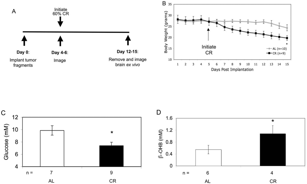
(Most of this is self-explanatory, but figure D is measuring ketone levels. Click to enlarge.)


The mice were induced with the brain cancer, and then supplied food either ad libitum (as much as they cared to eat) or at 40% of their normal intake once per day. The mice were measured for body weight, blood glucose levels, and ketone levels, as well as several different tests for the location and abundance of tumor cells. The calorie restricted (CR) mice had lower body weights, lower plasma glucose levels, and higher ketone levels than the ad libitum (AL) mice, as expected.


As you can see above, the cancer cells (dark purple) are spreading further and more diffuse into the cortex in the AL mice than the CR mice. And as you can see below, the number of cancer cells in a given histological section of the brain were higher in AL than CR mice.


Finally, one very interesting finding is that the CR mice also experienced decreased angiogenesis (generation of blood vessels) in tumor areas than AL mice. Both density and diameter of blood vessels was reduced in CR mice.


As it seems, caloric restriction decreases tumor spread and angiogenesis in a malignant brain cancer mouse model. This is strong evidence that cancer cells have difficulty proliferating under metabolic stress, even when healthy cells are functioning normally. Metabolic control, where appropriate, can be a non-invasive treatment for benign and malignant brain tumors, although like all cancer treatments it isn't without significant risk. It is also worth mentioning that CR did not completely stop the spread of the malignant cancer, although it did slow it down significantly, so it is likely that CR would have to be used in tandem with other treatments.


Shelton, L., Huysentruyt, L., Mukherjee, P., & Seyfried, T. (2010). Calorie restriction as an anti-invasive therapy for malignant brain cancer in the VM mouse ASN NEURO, 2 (3), 171-177 DOI: 10.1042/AN20100002